Anatomy Diagram Rib Area - Real Human Rib Thoracic Cage And Spine Bones Anatomy White 01 3d Model 119 Max Free3d - We also discuss the medical conditions and injuries that can affect these joints.
bymamamonnet•
0
Anatomy Diagram Rib Area - Real Human Rib Thoracic Cage And Spine Bones Anatomy White 01 3d Model 119 Max Free3d - We also discuss the medical conditions and injuries that can affect these joints.. We describe a minimally invasive laparoscopic approach to rib plating. They are twelve in number on either side; The primary responsibilities of the ribcage involve protecting the thoracic visceral organs, enclosing the thoracic visceral organs, and is included in the general mechanics of the process of this diagram with labels depicts and explains the details of rib cage anatomy. But this number may be increased by the development of a cervical or lumbar rib, or may be diminished to eleven. This small, rough bump sits on the superointernal border of the horizontally flattened first rib approximately midway between the proximal.
Lateral interchondral ligament of right seventh and eighth ribs. For more anatomy content please follow us and visit our website: This small, rough bump sits on the superointernal border of the horizontally flattened first rib approximately midway between the proximal. Includes images, video, and free quiz. Start studying anatomy of the rib.
Human Rib Cage High Res Stock Images Shutterstock from image.shutterstock.com Epidemiology associations rib fractures are often associated with other injuries and the greater the number of rib fractures the more likely are ass. This article looks at their anatomy and function and includes an interactive diagram. They extend from the lateral border of the costal grooves to the superior margins of the ribs below. The current morbidity of rib plating is due to the size of the incision required to perform an open procedure. All are attached at the back to the thoracic vertebrae and are numbered from 112 according to the vertebrae they attach to. By printing out this quiz and taking it with pen and paper creates for a. Human brain functional infographic diagram. We describe a minimally invasive laparoscopic approach to rib plating.
All are attached at the back to the thoracic vertebrae and are numbered from 112 according to the vertebrae they attach to.
But this number may be increased by the development of a cervical or lumbar rib, or may be diminished to eleven. Rib number 10 is atypical because its head. The first seven are connected behind with the vertebral column and in front. It has a roughened area on its upper surface, from which the serratus anterior muscle originates. Includes images, video, and free quiz. Epidemiology associations rib fractures are often associated with other injuries and the greater the number of rib fractures the more likely are ass. They extend from the lateral border of the costal grooves to the superior margins of the ribs below. They also have a role in. Instant anatomy is a specialised web site for you to learn all about human anatomy of the body with diagrams, podcasts and revision questions. In most tetrapods, ribs surround the chest, enabling the lungs to expand and thus facilitate breathing by expanding the chest cavity. The rib cage, shaped in a mild cone shape and more flexible than most bone sets, is made up of varying elements such as the thoracic vertebra, 12 equally paired ribs, costal cartilage, and held together anteriorly by the sternum. Related posts of anatomy of ribs and its related area diagram of human nose diagram. Lessons on the bone markings of the ribs and sternum.
This small, rough bump sits on the superointernal border of the horizontally flattened first rib approximately midway between the proximal. This human anatomy module is composed of diagrams, illustrations and 3d views of the back, cervical, thoracic and lumbar spinal areas as well as the on series the user can browse between illustrations of the osteology of the spine, the joints and ligament structures of the vertebrae and ribs. They extend from the lateral border of the costal grooves to the superior margins of the ribs below. The first seven are connected behind with the vertebral column and in front. Anatomy of the human rib cage.
Real Human Rib Thoracic Cage And Spine Bones Anatomy White 01 3d Model 119 Max Free3d from preview.free3d.com The ribs are elastic arches of bone, which form a large part of the thoracic skeleton. For more anatomy content please follow us and visit our website: In vertebrate anatomy, ribs (latin: This is a preview video for our tutorial about the anatomy of the ribs, the different types, their location and bony landmarks. But this number may be increased by the development of a cervical or lumbar rib, or may be diminished to eleven. Includes images, video, and free quiz. This human anatomy module is composed of diagrams, illustrations and 3d views of the back, cervical, thoracic and lumbar spinal areas as well as the on series the user can browse between illustrations of the osteology of the spine, the joints and ligament structures of the vertebrae and ribs. The skull and rib cage.
Learn vocabulary, terms and more with flashcards, games and other study tools.
Start studying anatomy of the rib. Interactive tutorials about the ribs and sternum bones, with labeled images and diagrams featuring the beautiful illustrations of getbodysmart. The first seven are connected behind with the vertebral column. We also discuss the medical conditions and injuries that can affect these joints. The first seven are connected behind with the vertebral column and in front. Learn everything about the ribs with our articles, video tutorials, quizzes, and labeled diagrams there are eleven pairs of external intercostal muscles and these are the most superficial in the area. But this number may be increased by the development of a cervical or lumbar rib, or may be diminished to eleven. In vertebrate anatomy, ribs (latin: Spare ribs diagram ribs anatomy types ossification u0026 clinical significance rib cage anatomy labeled vector illustration diagram 20.10.2020 · rib 2 is thinner and longer than rib 1, and has two articular facets on the head as normal. Anatomy of the human rib cage. Includes images, video, and free quiz. Human breathing, lung capacities, and breathing cycles.
The current morbidity of rib plating is due to the size of the incision required to perform an open procedure. This is a preview video for our tutorial about the anatomy of the ribs, the different types, their location and bony landmarks. They are twelve in number on either side; Includes images, video, and free quiz. They extend from the lateral border of the costal grooves to the superior margins of the ribs below.
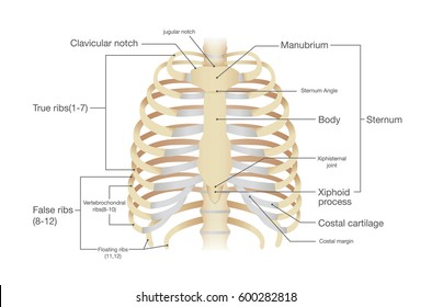
The Thoracic Cage The Ribs And Sternum Human Anatomy And Physiology Lab Bsb 141 from s3-us-west-2.amazonaws.com This article looks at their anatomy and function and includes an interactive diagram. It has a roughened area on its upper surface, from which the serratus anterior muscle originates. This image displays rib cage diagram. The first seven are connected behind with the vertebral column. They articulate with the vertebral column posteriorly, and terminate anteriorly as cartilage (known as costal cartilage). Lateral interchondral ligament of right seventh and eighth ribs. Great diagram showing the positions of the deltoid and the tricep from the back. Start studying anatomy of the rib.
Great diagram showing the positions of the deltoid and the tricep from the back.
Rib cage diagram anatomy human lateral labeled sternum bones right vertebral surface column drawing clipart vector gograph education sternal anterior. Rib number 10 is atypical because its head. The ribs are a set of twelve paired bones which form the protective 'cage' of the thorax. Lessons on the bone markings of the ribs and sternum. Spare ribs diagram ribs anatomy types ossification u0026 clinical significance rib cage anatomy labeled vector illustration diagram In vertebrate anatomy, ribs (latin: This image displays rib cage diagram. They articulate with the vertebral column posteriorly, and terminate anteriorly as cartilage (known as costal cartilage). Area between the head and the tubercle of the rib. 20.10.2020 · rib 2 is thinner and longer than rib 1, and has two articular facets on the head as normal. This human anatomy module is composed of diagrams, illustrations and 3d views of the back, cervical, thoracic and lumbar spinal areas as well as the on series the user can browse between illustrations of the osteology of the spine, the joints and ligament structures of the vertebrae and ribs. For more anatomy content please follow us and visit our website: Related posts of anatomy of ribs and its related area diagram of human nose diagram.